CT scan
NEOSCAN N60
COMPACT DESK-TOP X-RAY MICROTOMOGRAPH
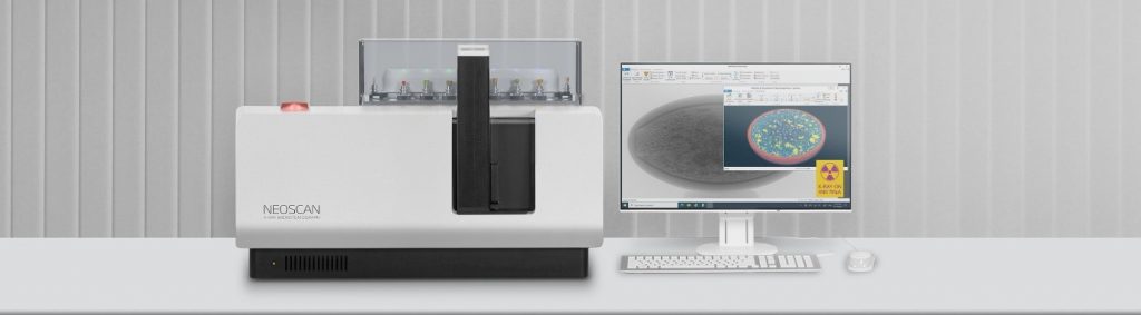
NEOSCAN N60 is a compact, desk-top, general purpose X-ray laboratory instrument for non-destructive 3-dimensional reconstruction of internal microstructure of the objects. Typical applications included, but not limited, by small molded or 3D printed parts in plastic, glass-fiber composites with 20-100µm fiber thickness, biological materials (tissue, small bones, etc.), entomology, food, small electronic assemblies and devices, geological samples (<10 mm), pharmaceutical products and packaging (tablets, pills, capsules, …), etc.
SPECIFICATIONS:
General: | |
Pixel size at maximum magnification | < 5 µm |
True low-contrast 3D resolution | 10 µm or better |
Maximum scanning diameter (single scan/offset scan) | 15 mm / 30 mm |
Maximum scanning length | 60 mm |
Maximum physical object length | 100 mm |
Radiation safety | < 1µSv/h at 10cm from instrument surface |
Size / weight | 800W x 310D x 375H mm / 65kg |
Power supply | 100 – 240 V AC, 50 – 60 Hz, 3A |
X-Ray Source: | |
Type | metal-ceramic, permanently sealed |
Emitter | W, reflective target |
Maximum voltage | 65 kV |
Maximum power | 50 W |
Automatic filter changer, number of positions | 4 |
X-Ray Detector: | |
Image sensor | 8 megapixels, cooled |
Camera field of view | 18 x 13 mm |
Scintillator | GADOX |
Fibre-optics | 1 : 1, radiation hardened |
The NEOSCAN N60 based on newly developed key components, such as advanced metal-ceramic microfocus X-ray source and specially designed for this system large-format X-ray detector. It also includes precision electromechanical manipulator controlled by new electronics with micro-stepping drives, and integrated software package for scanning, reconstruction and visualization of obtained results.
The X-ray source is a permanently sealed, maintenance-free metal-ceramic tube with big power and extremely long lifetime. An acquisition geometry used in NEOSCAN N60 with object positioned close to detector and spatial resolution fully defined by detector allows using X-ray source with significantly high power than in other microtomography instruments without compromising spatial resolution.
The X-ray detector based on cooled 8-megapixel imaging sensor directly attached to radiation-hardened fiber-optic plate coated by directly deposed scintillator. Small pixel size in image sensor and specially produced fiber-optics with very thin, radiation-protective fibers ensure high image quality and long lifetime without radiation damages.
Specially designed object manipulator in combination with drive electronics for micro-stepping control of all motors provides micron-level accuracy in object positioning and rotation during scanning.
The NEOSCAN N60 system includes internal shielding sufficient for using this instrument in standard laboratory environments according to all safety regulations without any additional requirements to installation areas.
The system supplied with variety of stages for different object sizes.
The system can be supplied with optional computer with all preinstalled software, or with software package on flash drive for installation on customer computer. The system requires one USB3 connection to control computer. Minimum computer configuration is following:
- Laptop or desktop with available USB3 port
- 1GHz or faster Intel processor
- 2GB or more memory
- 100GB or more hard drive
- NVidia graphics recommended
- Microsoft Windows-10 Pro
Supplied software includes integrated package for Scanning / Reconstruction / Visualization with friendly Microsoft-Office style user interface.
Optional Parts:
The NEOSCAN N60 can be supplied with optional Computer in following configuration with all preinstalled software:
Microsoft Surface Book 2 or similar:
- Intel Core i7 processor
- 16GB memory
- 512GB Solid State Drive
- NVidia GTX1060 integrated graphics
- 15” touchscreen with native 3240×2160 pixels
Service and Support:
The NEOSCAN N60 system supplied with standard 12 months warranty. Extended warranty can be purchased separately.
For service and training purposes, it is strongly recommended to get good internet connection in the computer used for system control.
NEOSCAN N80(CCD)
DESK-TOP HIGH RESOLUTION X-RAY MICROTOMOGRAPH
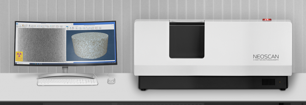
NEOSCAN N80 is a desk-top, general purpose X-ray laboratory instrument for non-destructive 3-dimensional reconstruction of internal microstructure of the objects with spatial resolution in micron range. Typical applications include, but not limited, a medium-large size plastic molded or 3D printed parts, light metals (Al, Ti) and small parts in more dense metals (steel, …), ceramics, carbon- and glass-fiber composites, biological materials (tissue samples, bones, implants, …), electronics, glass, geological samples – plugs and drilled cores, pharmaceutical products and packaging, etc.
SPECIFICATIONS:
General: | |
Pixel size at maximum magnification | < 0,5 µm |
True low-contrast 3D resolution | 2 µm or better |
Maximum scanning diameter (single scan/offset scan) | 50 mm / 100 mm |
Maximum scanning length | 130 mm |
Maximum physical object length | 200 mm |
Radiation safety | < 1µSv/h at 10cm from instrument surface |
Size / weight | 1260W x 638D x 510H mm / 260kg |
Power supply | 100 – 240 V AC, 50 – 60 Hz, 3A |
X-Ray Source: | |
Type | permanently sealed |
Emitter | W,transmission target |
Maximum voltage | 110kV |
Maximum power | 16W |
Smallest spot size | 2 µm or smaller |
Automatic filter changer, number of positions | 20 |
X-Ray Detector: | |
Image sensor | 16 megapixels, cooled |
Camera field of view | 72 x 48 mm |
Scintillator | GADOX |
Fibre-optics | 2 : 1 |
Protection against radiation damages | radiation hardened |
Integrated options: | |
Micro-positioning stage (travel) | Integrated (10 mm) |
Power for optional stages through sliprings | 24 W (24 V / 1 A) |
Shielded passthrough for additional cables/tubes | Integrated |
The NEOSCAN N80 based on newly developed key components, such as microfocus permanently sealed X-ray source and large-format X-ray detector, with specially designed precision electromechanical manipulator, new electronics with micro-stepping drives, and integrated software package for scanning, reconstruction and visualization of obtained results.
The X-ray source has a permanently sealed, maintenance-free construction with unique 2 micrometer spot size, which provides very high spatial resolution of the system. Possibility for target repositioning by operator ensure extremely long lifetime of the source. Using transmission type target allows placing objects very close to emission point and correspondingly reduce distance between the source and X-ray detector with corresponding reduction of scanning time and improvement in signal-to-noise ratio.
The X-ray detector contains large format cooled imaging sensor directly attached to 2:1 fiber-optic taper covered by radiation hardened fiber-optic plate coated by directly deposed scintillator. Such construction with dual fiber-optic design allows 4-fold increase of field of view and ensure strong resistance against radiation damages. Large field of view allows scanning big samples and collecting 4 times more X-ray radiation emitted by X-ray source with corresponding reduction of scanning time and improvement of image quality.
New electromechanical object manipulator in combination with drive electronics for micro-stepping control of all motors provides micron-level accuracy in object positioning and rotation during scanning. The system includes integrated micro-positioning stage on the top of object rotation assembly for exact sample positioning or for selection of scanning volume of interest inside big samples.
The NEOSCAN N80 system includes internal shielding against radiation leaks sufficient for using this instrument in standard laboratory environment according to all safety regulations without any additional requirements to installation areas. Due to significant weight, the system should be installed on strong, stable desk (not supplied with the system) in vibration-free room.
The system supplied with variety of stages for different object sizes including special stage for large objects with three independently adjusted jaws.
The system can be supplied with optional workstation in one of two standard configurations with all preinstalled software, or with software package on flash drive for installation on customer computer. The system requires one USB2 connection and one FireWire (6-ways) connection to control computer. Minimum computer configuration is following:
- Intel XEON Bronze or Intel Core i7 processor, 2GHz or faster,
- 32GB memory,
- 1TB hard drive or solid-state drive,
- NVIDIA Quadro P1000 or RTX4000 graphics card,
- IEEE1394 (FireWire) card with 6-pin connector,
- One available USB2 or USB3 port,
- QWERTY keyboard, mouse,
- 21 inch or larger HD monitor,
- Windows 10 Pro.
Supplied software includes integrated package for Scanning / Reconstruction / Visualization with friendly Microsoft-Office style user interface.
Optional Parts:
- In the case of using system in laboratories where possible vibrations can occur, the system can be equipped with optional pneumatic vibration isolators (should be ordered with the system, factory installed before system delivery). The set of vibration isolators contains four pneumatic feet installed to the corners of the system body. The feet with very low resonance frequency allow effective rejection of vibrations coming from the surface where the system will be installed.
- The NEOSCAN N80 can be supplied with optional Workstation with all preinstalled software in one of two following configurations:
“Standard” version: DELL Precision 7820 or similar:
- Intel XEON Silver 4208, 2.1/3.2GHz, 8-core processor,
- NVIDIA Quadro P2000, 5GB graphical card,
- 48GB RAM,
- 512GB Solid State Drive for OS and programs,
- 8TB, 7200rpm SATA HDD,
- IEEE1394 (FireWire) card with 6-pin connector,
- QWERTY keyboard, optical mouse,
- 24-inch UltraSharp LCD monitor, 1920×1200 native resolution,
- Windows 10 Pro.
“Advanced” version: DELL Precision 7920 or similar:
- Two Intel XEON Gold 5218, 2.3/3.9GHz processors, 16 cores each,
- NVIDIA Quadro P5000, 16GB graphical card,
- 128GB RAM,
- 1TB Solid State Drive for OS and programs,
- 16TB (2x8TB), 7200rpm SATA HDD,
- IEEE1394 (FireWire) card with 6-pin connector,
- QWERTY keyboard, optical mouse,
- 34-inch Ultra-Wide Curved LCD monitor, 3440×1440 native resolution,
- Windows 10 Pro.
Service and Support:
The NEOSCAN N80 system supplied with standard 12 months warranty. Extended warranty can be purchased separately.
For service and training purposes, it is strongly recommended to get good internet connection in the computer used for system control.
NEOSCAN N80(FP)
DESK-TOP HIGH RESOLUTION X-RAY MICROTOMOGRAPH
NEOSCAN N80 is a desk-top, general purpose X-ray laboratory instrument for non-destructive 3-dimensional reconstruction of internal microstructure of the objects with spatial resolution in micron range. Typical applications include, but not limited, a medium-large size plastic molded or 3D printed parts, light metals (Al, Ti) and small parts in more dense metals (steel, …), ceramics, carbon- and glass-fiber composites, biological materials (tissue samples, bones, implants, …), electronics, glass, geological samples – plugs and drilled cores, pharmaceutical products and packaging, etc.
SPECIFICATIONS:
General: | |
Pixel size at maximum magnification | < 1 µm |
True low-contrast 3D resolution | 2 µm or better |
Maximum scanning diameter (single scan/offset scan) | 50 mm / 100 mm |
Maximum scanning length | 130 mm |
Maximum physical object length | 200 mm |
Radiation safety | < 1µSv/h at 10cm from instrument surface |
Size / weight | 1260W x 638D x 510H mm / 225kg |
Power supply | 100 – 240 V AC, 50 – 60 Hz, 3A |
X-Ray Source: | |
Type | permanently sealed |
Emitter | W,transmission target |
Maximum voltage | 110kV |
Maximum power | 16W |
Smallest spot size | 2 µm or smaller |
Automatic filter changer, number of positions | 20 |
X-Ray Detector: | |
Image sensor | 7 megapixels, cooled |
Camera field of view | 140 x 120 mm |
Scintillator | GADOX |
Fibre-optics | 1 : 1 |
Protection against radiation damages | radiation hardened |
Integrated options: | |
Micro-positioning stage (travel) | Integrated (10 mm) |
Power for optional stages through sliprings | 24 W (24 V / 1 A) |
Shielded passthrough for additional cables/tubes | Integrated |
The NEOSCAN N80 based on newly developed key components, such as microfocus permanently sealed X-ray source and large-format X-ray detector, with specially designed precision electromechanical manipulator, new electronics with micro-stepping drives, and integrated software package for scanning, reconstruction and visualization of obtained results.
The X-ray source has a permanently sealed, maintenance-free construction with unique 2 micrometer spot size, which provides very high spatial resolution of the system. Possibility for target repositioning by operator ensure extremely long lifetime of the source. Using transmission type target allows placing objects very close to emission point and correspondingly reduce distance between the source and X-ray detector with corresponding reduction of scanning time and improvement in signal-to-noise ratio.
The X-ray detector contains large format cooled imaging sensor directly attached to 2:1 fiber-optic taper covered by radiation hardened fiber-optic plate coated by directly deposed scintillator. Such construction with dual fiber-optic design allows 4-fold increase of field of view and ensure strong resistance against radiation damages. Large field of view allows scanning big samples and collecting 4 times more X-ray radiation emitted by X-ray source with corresponding reduction of scanning time and improvement of image quality.
New electromechanical object manipulator in combination with drive electronics for micro-stepping control of all motors provides micron-level accuracy in object positioning and rotation during scanning. The system includes integrated micro-positioning stage on the top of object rotation assembly for exact sample positioning or for selection of scanning volume of interest inside big samples.
The NEOSCAN N80 system includes internal shielding against radiation leaks sufficient for using this instrument in standard laboratory environment according to all safety regulations without any additional requirements to installation areas. Due to significant weight, the system should be installed on strong, stable desk (not supplied with the system) in vibration-free room.
The system supplied with variety of stages for different object sizes including special stage for large objects with three independently adjusted jaws.
The system can be supplied with optional workstation in one of two standard configurations with all preinstalled software, or with software package on flash drive for installation on customer computer. The system requires one USB2 connection and one FireWire (6-ways) connection to control computer. Minimum computer configuration is following:
- Intel XEON Bronze or Intel Core i7 processor, 2GHz or faster,
- 32GB memory,
- 1TB hard drive or solid-state drive,
- NVIDIA Quadro P1000 or RTX4000 graphics card,
- IEEE1394 (FireWire) card with 6-pin connector,
- One available USB2 or USB3 port,
- QWERTY keyboard, mouse,
- 21 inch or larger HD monitor,
- Windows 10 Pro.
Supplied software includes integrated package for Scanning / Reconstruction / Visualization with friendly Microsoft-Office style user interface.
Optional Parts:
- In the case of using system in laboratories where possible vibrations can occur, the system can be equipped with optional pneumatic vibration isolators (should be ordered with the system, factory installed before system delivery). The set of vibration isolators contains four pneumatic feet installed to the corners of the system body. The feet with very low resonance frequency allow effective rejection of vibrations coming from the surface where the system will be installed.
- The NEOSCAN N80 can be supplied with optional Workstation with all preinstalled software in one of two following configurations:
“Standard” version: DELL Precision 7820 or similar:
- Intel XEON Silver 4208, 2.1/3.2GHz, 8-core processor,
- NVIDIA Quadro P2000, 5GB graphical card,
- 48GB RAM,
- 512GB Solid State Drive for OS and programs,
- 8TB, 7200rpm SATA HDD,
- IEEE1394 (FireWire) card with 6-pin connector,
- QWERTY keyboard, optical mouse,
- 24-inch UltraSharp LCD monitor, 1920×1200 native resolution,
- Windows 10 Pro.
“Advanced” version: DELL Precision 7920 or similar:
- Two Intel XEON Gold 5218, 2.3/3.9GHz processors, 16 cores each,
- NVIDIA Quadro P5000, 16GB graphical card,
- 128GB RAM,
- 1TB Solid State Drive for OS and programs,
- 16TB (2x8TB), 7200rpm SATA HDD,
- IEEE1394 (FireWire) card with 6-pin connector,
- QWERTY keyboard, optical mouse,
- 34-inch Ultra-Wide Curved LCD monitor, 3440×1440 native resolution,
- Windows 10 Pro.
Service and Support:
The NEOSCAN N80 system supplied with standard 12 months warranty. Extended warranty can be purchased separately.
For service and training purposes, it is strongly recommended to get good internet connection in the computer used for system control.
Software NeoScan
Supplied software includes integrated package for Scanning / Reconstruction / Visualization with friendly Microsoft-Office style user interface. One of three possible modes can be selected by top tabs.
Scanning Mode:
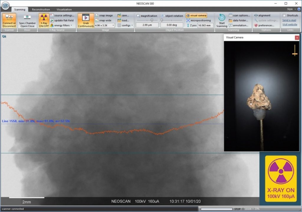
The Scanning Mode allows initializing the scanner, provides access to specimen chamber for object installation or replacement, activates / deactivates X-ray source, adjusts (manually or automatically) source parameters and energy filtering, collects image from X-ray detector, performs all image corrections including active artifact elimination, saves/loads single image or sequence of images during object rotation for following tomographical reconstruction, adjusts object position and image magnification, shows visual image of the object inside specimen chamber using internal color optical camera, performs all calibration procedures and sets / applies system settings. It can store full set of system settings as a user configuration to restore system set-up for specific study on request. An operator can measure object dimensions and show intensity profiles on the screen by simple mouse clicks. The object can be positioned in the specimen chamber by dragging image with mouse on the screen or by numerical values for pixel size, rotation angle and elevation. Long objects can be scanned by automatically performed multiple partial scans, which will be connected to single set of virtual slices through the object during following reconstruction.
The software includes intensive monitoring of the system with measurements of rotation speed for all four internal cooling fans, measuring cooling temperature of imaging sensor inside X-ray detector, reading parameters and operation time of X-ray source, supply voltages for all key components, etc.
Reconstruction Mode:
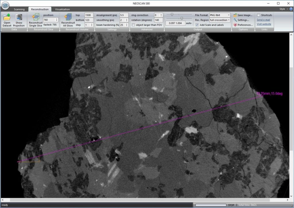
The Reconstruction Mode allows converting set of two-dimensional images of the object acquired during scanning to a stack of virtual slices through the object to reveal its internal microstructure non-destructively. It works with intensive multithreading using all available cores of computer processors. An operator can reconstruct any single, multiple or all virtual cross sections through the object. A gray-scale window for all resulting cross section can be selected manually or automatically. The software can perform automatic misalignment correction, apply smoothing to reduce noise on reconstructed slices, add “beam hardening correction” to compensate impact of different absorption for different X-ray energies in polychromatic primary beam, apply ring artifact correction, implement reconstruction with selected slice rotation angle and reconstruct objects larger than field of view. All results can be saved in conventional image formats, such as 16-bit TIFF, 8-bit BMP, JPG or PNG. The reconstruction can be performed in full scanning volume or in selected region of interest with square, rectangular, circular or elliptical shape selected in size and position by operator. Advanced settings allow adjusting spectral window of convolution procedure involved in reconstruction process and limit processor load to allow performing other tasks during reconstruction process.
The reconstructed slices can be saved to local or remote (network) location with possibility for automatic creation of subfolders with incremental names and adding incremental suffixes to the names of resulting set of cross sections.
Visualization Mode:
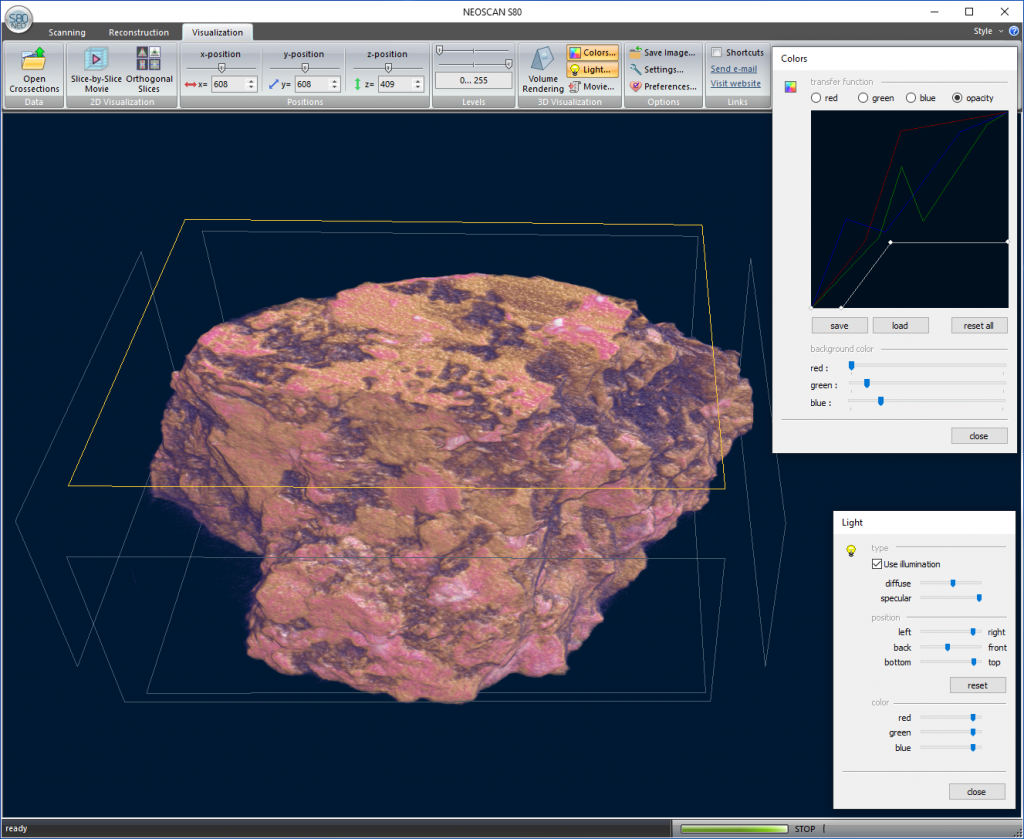
The Visualization Mode can work in two possible approaches: visualization of two-dimensional images or volume rendering for realistic displaying of full three-dimensional structure. Two-dimensional visualization can display reconstructed results as a slice-by-slice movie or as three orthogonal slices through the objects crossed at any point or reconstructed volume. An operator can select any position of cross point and adjust grayscale levels of displayed absorption range. In volume rendering mode, one can see full reconstructed object as a realistic three-dimensional image with possibility to zoom in and out, rotate, make virtual cut, manipulate clipping cube and object volume separately or together, select color and transparency curves, add illumination with controllable direction and color, adjust viewing angle, etc. Integrated “movie creator” allows creating movies with flights around or inside the object by simple selection of several key frames with following automatic interpolation of flying trajectory and all color / transparency / light properties for all intermediate frames. The final movie with selected framerate will be saved in efficiently compressed MP4 file, which can be played by double click using integrated in Windows players. Any screen image in any visualization mode can be also saved as BMP, JPG or PNG file.